The Future of ccRCC Imaging
The Future of ccRCC Imaging
CAIX imaging could offer accurate, noninvasive diagnosis of ccRCC
Existing imaging modalities fall short in their ability to distinguish clear cell renal cell carcinoma (ccRCC) from other renal tumors, causing diagnostic uncertainty.1 However, a validated glycoprotein target called carbonic anhydrase IX (CAIX or CA9) is poised to set a new standard for renal mass evaluation.2
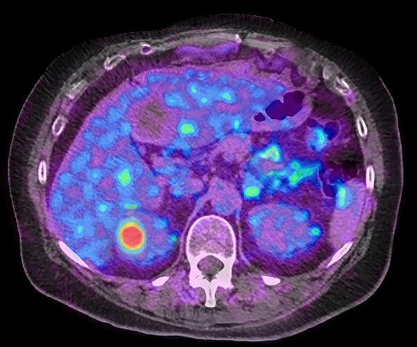
CAIX imaging aims to revolutionize the management of ccRCC, just as PSMA PET changed the management of prostate cancer.
Case study: 1.8-cm renal mass, unknown biology
Medical history
- 70-year-old male patient with well-controlled hypertension presented with GI-related discomfort
- Patient received abdominal CT which revealed a 1.8-cm lesion on his left kidney
Click each tab to view potential outcomes


Given the lesion’s small size and the patient’s lack of symptoms, renal mass biopsy is not performed. The patient expresses strong aversion to surgery, leading to initiation of active surveillance. He is closely monitored with periodic imaging every 6 months, with a focus on lesion growth rate and emergence of new symptoms.
After 3 years of surveillance, tumor growth leads to intervention.


The patient is offered noninvasive CAIX imaging, a novel molecular scan targeting ccRCC—the most common RCC subtype.4 The scan yields positive, high-contrast uptake in the lesion, indicative of ccRCC.
Armed with greater confidence about the tumor biology, the patient and his care team proceed with an appropriate treatment plan.

By clarifying tumor biology up front, care teams can create individualized treatment plans, potentially improving overall outcomes for their patients.5
Shining New Light on Diagnostic Precision in ccRCC
89Zr-girentuximab, an investigational radiodiagnostic imaging agent for the diagnosis and characterization of renal masses as ccRCC, received Breakthrough Designation.6
Having 89Zr included in your license may help shorten patients’ time to an accurate ccRCC diagnosis
Ensuring your radioactive material license includes 89Zr will allow for timely access to this noninvasive diagnostic option.
References: 1. van Oostenbrugge TJ, Fütterer JJ, Mulders PFA. Diagnostic imaging for solid renal tumors: a pictorial review. Kidney Cancer. 2018;2(2):79-93. 2. Stillebroer AB, Mulders PFA, Boerman OC, Oyen WJG, Oosterwijk E. Carbonic anhydrase IX in renal cell carcinoma: implications for prognosis, diagnosis, and therapy. Eur Urol. 2010;58(1):75-83. 3. Data on file. Telix Pharmaceuticals, Ltd. 4. Qi X, Li Q, Che X, Wang Q, Wu G. The uniqueness of clear cell renal cell carcinoma: summary of the process and abnormality of glucose metabolism and lipid metabolism in ccRCC. Front Oncol. 2021;11:727778. 5. Abou Elkassem AM, Lo SS, Gunn AJ, et al. Role of imaging in renal cell carcinoma: a multidisciplinary perspective. Radiographics. 2021;41(5):1387-1407. 6. FDA accepts BLA for TLX250-CDx (Zircaix®) for kidney cancer imaging, grants priority review. News release. Telix Pharmaceuticals. February 26, 2025. Accessed October 27, 2025. https://telixpharma.com/news-views/fda-accepts-bla-for-tlx250-cdx-zircaix-for-kidney-cancer-imaging-grants-priority-review/